Syringoma vs Milia: Depth, Look, Recurrence and the Right Treatment
Both look like tiny bumps under the eyes, but one sits at the surface and one sits deep in the dermis. A dermatologist explains how to tell them apart.

Whether this treatment suits you needs a dermatologist to assess. Send a photo and your travel dates on WhatsApp, and we reply in English whether it can be arranged for your case and which days are open.
Ask us on WhatsApp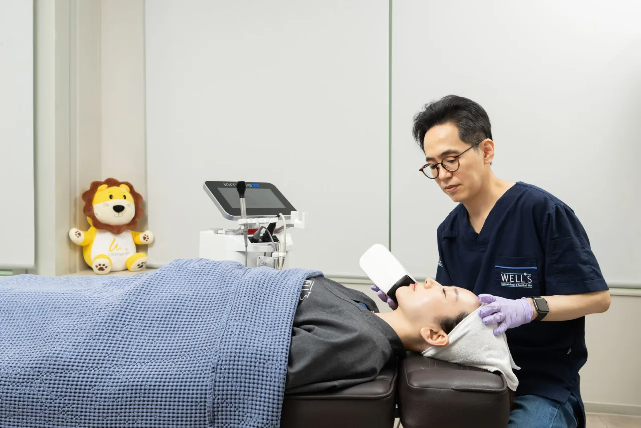
Syringoma vs milia: the short answer
Hello, I'm Dr. Won Sin Lee, a board-certified dermatologist and Chief Director at WELLS Clinic in Apgujeong, Seoul. I trained and was board certified at Asan Medical Center, where I am an adjunct professor of dermatology, and I co-founded WELLS in 2008. Patients often ask me about syringoma vs milia, usually after months of treating small under-eye bumps that never changed.
Here is the answer first. Milia sit in the epidermis; syringoma sits deep in the dermis. Milia are a little ball of keratin trapped just under the surface, and they can be opened and cleared. Syringoma is a growth of sweat-duct tissue that reaches the lower dermis, resembles milia, and recurs easily, so it needs a dermatologist's diagnosis and a deeper treatment.
Everything else in this post follows from that difference in depth: how each one looks, why squeezing works for one and harms the other, and what treatment actually reaches the problem.
| Milia | Syringoma | |
|---|---|---|
| What it is | Keratin that failed to shed and stayed lodged | Proliferation of sweat-gland ducts |
| Depth | Epidermis (surface layer) | Reaches the lower dermis |
| Typical look | Firm, white, pearl-like dot | Skin-colored, slightly raised, often shiny |
| Pattern | Single or a few scattered dots | Often clusters, frequently on both lower eyelids |
| Squeezing at home | Technically possible, but risks infection and scarring | Not possible; forcing it can inflame or pigment the skin |
| Coming back | A cleared milium is gone, new ones can form | Recurs easily |
Worried about your own case? Let a dermatologist look first: send a photo and your dates, and we reply whether it can be arranged.
Ask us on WhatsAppHow can you tell syringoma from milia in the mirror?
Colour is the most useful first clue. Milia look like a tiny grain of white keratin sitting under a thin skin cover. Syringoma is usually the colour of the surrounding skin, sometimes faintly yellowish, and has a slight sheen when light catches it.
Shape and texture differ too. A milium is a small, round, firm dome. Syringomas tend to be flatter on top and to sit in groups, so the area looks gently bumpy rather than dotted.
Milia under the eyes vs syringoma around the eyes
Both love the same real estate, which is why the confusion exists. Milia under the eye or on the eyelid are common, and they can appear at any age. Syringomas are classically found on the lower eyelids, often roughly symmetrical on both sides, and in some people they also appear on the cheeks, forehead, neck or chest.
- Leans milia: a few bright white, hard dots that look like they have a seed inside.
- Leans syringoma: many small skin-coloured bumps in a band along both lower lids.
- Either way: if you are not sure, you are in good company, because these are genuinely hard for non-specialists to tell apart.

I am careful not to diagnose from a mirror description, and you should be careful too. In the clinic we look at the bumps in good light and under magnification, which settles most cases quickly.
Why depth decides everything
Skin has two main layers. The epidermis is the outer layer that makes and sheds keratin; the dermis lies below it and holds structures such as sebaceous glands and sweat glands.
Milia form in the epidermis, so a dermatologist can reach and remove them with very little disturbance to the skin around them. Syringoma comes from the sweat-gland ducts in the dermis and extends down to the lower dermis. There is nothing inside a syringoma to squeeze out, because the bump is the tissue itself.
That is why the home approaches people try for milia rarely touch syringoma. Exfoliating acids, scrubs and rich eye creams work on the surface, and the structure that makes a syringoma lies well below it.
Can I squeeze milia, or syringoma, at home?
Because milia are confined to the epidermis, they can technically be extracted. I still recommend having it done at a dermatology clinic, since a needle at home and thin under-eye skin add up to a real risk of infection and scarring.
Syringoma cannot be extracted at home at all. Forcing it tends to cause inflammation or pigmentation that can last longer than the bump would have bothered you. My longer guide on how to get rid of milia under eyes covers the home-care question in more detail.
Not sure which one you have? Send a clear, well-lit photo of the area and your travel dates, and we'll reply with what we think we're looking at and how a visit would work.
Send a photo on WhatsAppWhy does syringoma keep coming back?
This is the part of syringoma vs milia that matters most for expectations. A milium that has been properly cleared is gone, although new ones can form elsewhere. Syringoma is different: it recurs easily, and our warts and syringoma page says so plainly.
Part of the reason is depth. Treatment has to reach deep tissue without wrecking the surface, so a cautious session can leave some ductal tissue behind. Part of it is biology: syringoma is linked with sweat ducts proliferating as skin ages, so new bumps can appear over time.
Two other questions come up in almost every consultation. Syringoma is not contagious and does not spread from one bump to another. Whether it is hereditary is not clearly established, though similar patterns sometimes appear within a family that shares skin traits and habits.
When the diagnosis needs a closer look
Syringoma is hard to self-diagnose precisely because it resembles milia. Our clinic's position is that it should be confirmed histologically, meaning under the microscope when the clinical picture is not clear, and treated by an experienced dermatologist. That step protects you from months of treatment aimed at the wrong lesion.
How is each one treated?
For both bumps the aim is the same: remove the structure itself, at the depth where it sits, without damaging the skin around it. The difference is how deep that is.
Milia: a surface problem
Because milia are superficial, removing them is generally the simpler of the two. The skill lies in stopping at the right depth rather than going deeper than the bump needs. If your bumps turn out to be milia, the milia under eyes guide walks through what clinic removal involves.
Syringoma: a deep problem
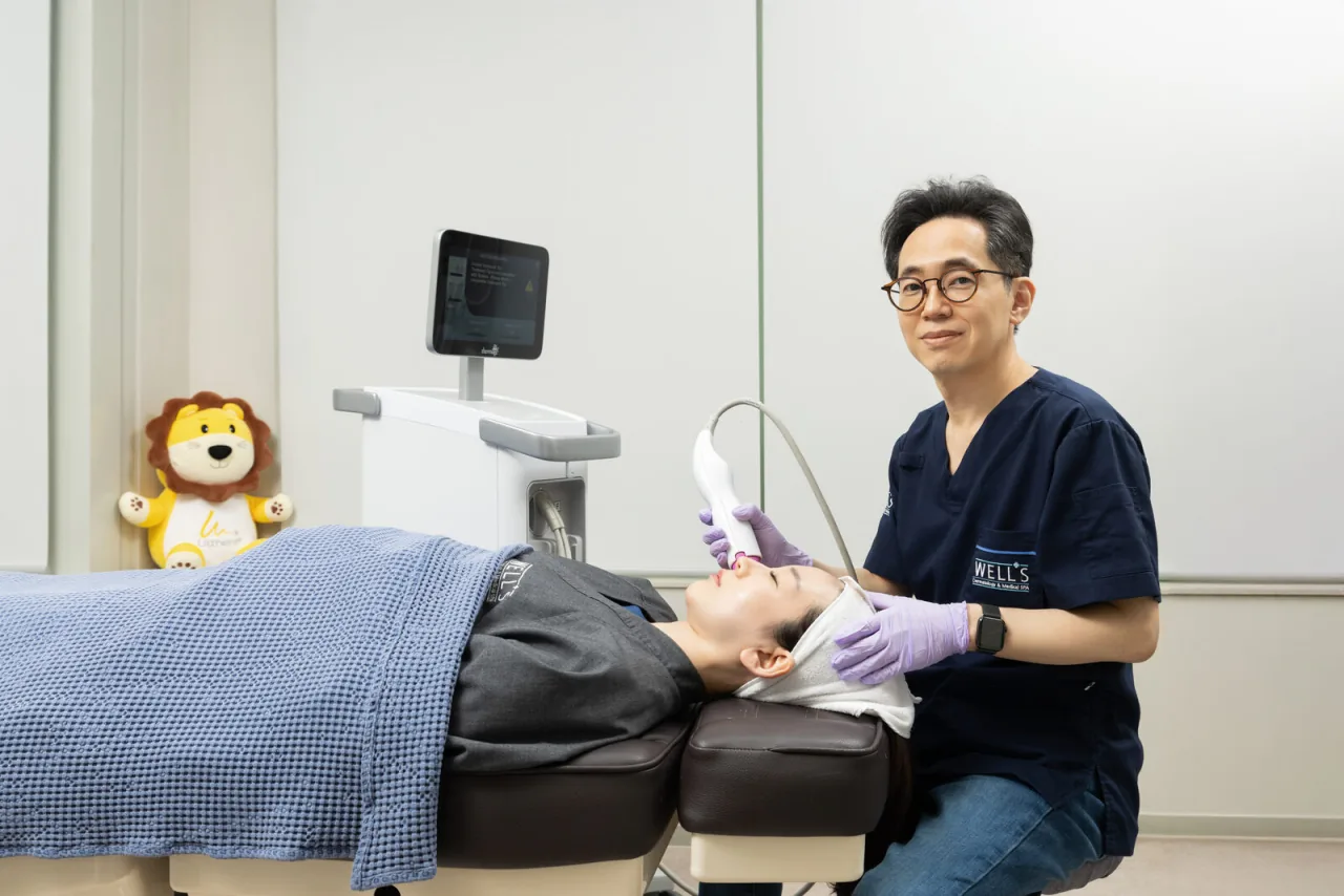
Syringoma needs an approach built for depth. Repeatedly ablating it from the surface with a CO2 laser easily leaves scars, because you have to burn through healthy upper skin to reach it. Insulated radiofrequency microneedling, which spares the surface while delivering energy to the deeper tissue, is generally the better choice.

How many sessions you need depends on how many bumps there are, how deep they sit and how your skin heals, so I discuss that at the consultation rather than promise a number in advance. The listed position for this treatment is simple: price confirmed at consultation.
What else looks like milia or syringoma?
Two more benign bumps join this line-up often enough to mention. Telling all four apart is why the first visit is about diagnosis, not about a device.
- Flat warts: slightly raised epidermis caused by a virus. They are the only one of the group that spreads, and picking them spreads the virus. Flat warts are often two to three times more numerous than you can see.
- Sebaceous hyperplasia: an overgrown oil gland in the upper dermis, skin-toned with a yellowish, oily tinge. A single ordinary laser pass tends to leave the gland behind, so it comes back.
- Closed comedones: small whiteheads that are a form of acne, not keratin cysts. If that is your question, see milia vs comedone.
On warts vs milia specifically: warts tend to be flat-topped and skin-coloured or light brown, and they multiply, while milia are white, round and stay put. Because warts and syringomas share a treatment page at WELLS, both are assessed in the same consultation.
Getting a syringoma or milia diagnosis at WELLS from abroad
At WELLS, consultation, diagnosis and treatment are done by a board-certified dermatologist from start to finish. That matters more for bumps like these than for almost anything else, because the diagnosis is the treatment plan.
- Message us on WhatsApp with your concern, a photo if you like, and your travel dates. We reply in English with options and available times.
- At the clinic, a dermatologist examines the skin, confirms which bump it is, and confirms the plan and price before anything starts.
- Treatment and aftercare happen in the same clinic, and you can reach us on WhatsApp afterwards.
We are on the 8th floor of the Apgujeong Building, 842 Nonhyeon-ro, Gangnam-gu, about five minutes on foot from Apgujeong Station, Line 3, Exit 3. You can read more about my background on my doctor profile, and about the other conditions we treat on the skin conditions page.
If you are planning a trip to Seoul, send your dates and a photo of the area. We'll tell you what the first visit would cover and how to fit it around your flights.
Ask about a consultationFind out which bump you have before you treat it
Send a clear photo of the area and your travel dates on WhatsApp. A dermatologist-led team will reply in English with what the consultation would cover and available times in Apgujeong.
Message the clinic on WhatsAppReplies on WhatsApp · prices in KRW, including 10% VATBefore you message us
Are syringoma and milia the same thing?
No. Milia are trapped keratin in the epidermis, the surface layer of skin. Syringoma is a proliferation of sweat-gland ducts that reaches the lower dermis. They can look alike under the eyes, but the depth and the treatment are different.
Can syringoma turn into something dangerous?
Syringoma is a benign growth and it is not contagious. The practical concern is that it is easy to mistake for milia or other bumps, so any bump that grows, bleeds, crusts or changes colour should be checked by a dermatologist.
Why did my under-eye bumps come back after treatment?
If they were syringoma, recurrence is common because the tissue sits deep in the dermis and the ducts can keep proliferating with skin ageing. If they were treated as milia, the depth may simply have been wrong. A dermatologist's diagnosis is the first step either way.
What is the difference between warts vs milia?
Flat warts are slightly raised epidermis caused by a virus; they tend to be flat-topped, skin-coloured and to multiply, and picking them spreads the virus. Milia are white, firm keratin dots that do not spread.
How much does syringoma treatment cost at WELLS?
The price for warts and syringoma treatment is confirmed at your consultation, once the dermatologist has seen how many bumps there are and how deep they sit. Prices at WELLS are quoted in Korean won with 10% VAT included.
Results and recovery vary by individual. Every treatment carries possible side effects, which are explained at the consultation. Prices are in Korean won, including 10% VAT, and confirmed at your consultation. This article is general information from the clinic, not a diagnosis.
Dr. Won Sin Lee
- Board-certified dermatologist in Korea; trained at Asan Medical Center in Seoul
- Dermatology residency, Asan Medical Center; board certification in dermatology in Korea
- Adjunct professor, Department of Dermatology, Asan Medical Center
- Former director, Bokji Dermatology Clinic, Korean Leprosy Welfare Association
- Commendation for Outstanding Service as a Public Health Physician